Global English
 UK
UK


This blog is the second in a series of three instalments, preceeded by Part 1 (Ethyology & Pathogenesis of the infection) and followed by Part 3 (Therapy & Treatment).
Pyoderma is a skin infection caused by pyogenic bacteria, usually Staphylococci, and is one of the most frequent diseases in veterinary dermatology. It is characterised by a pleomorphic clinical presentation, which can often mimic many other skin diseases such as demodicosis, dermatophytosis or pemphigus foliaceus. Rarely occurring as a primary form, in most cases it represents a complication of a pre-existing disease, such as atopy, food allergy, horny disorders, parasitic diseases, endocrinopathies and immunodeficiency states.
Clinical overview
Superficial pyoderma can have a variety of clinical pictures. Intertrigo (Pic. 1) is the superficial infection of the skin, deep in the skin folds, typical of certain breeds, such as Shar-Pei, French and English Bull Dogs and the boxer, or of the interdigital spaces, typical of many allergic dogs. Erythema and skin moisture with a whiteish exudate rich in bacteria on cytological examination are observed.
Picture 1 – Intertrigo on the muzzle of a brachycephalic dog
Pyotraumatic dermatitis (Pic. 2) is the superficial, particularly exudative and itchy infection of lesions caused by dog biting. These lesions are frequently associated with flea bite allergy and localised to the hindlimb.
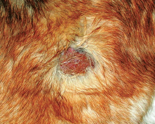
Picture 2 – Pyotraumatic dermatitis on the lateral thigh of a dog with flea allergy
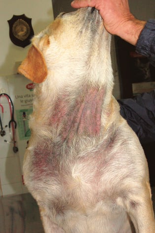
The "bacterial overgrowth" (Pic. 3) is characterised by large skin areas, often ventral and medial (armpits, groin, ventral neck) characterised by alopecia, lichenification, hyperpigmentation and a surface damp to the touch and strongly smelling. On cytological examination, excessive numbers of bacteria are observed on the skin surface.
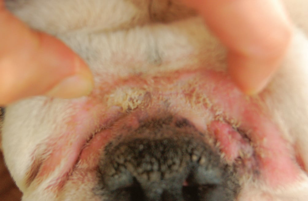
 Bacterial folliculitis (Pic. 4) is characterised by papules, pustules, which easily break off resulting in epidermal collarettes and focal alopecia. A particular form of bacterial folliculitis, called superficial spreading pyoderma, manifests itself without pustules, but with large collarets and an erythematous front at the margins. Pyoderma of the mucocutaneous junctions (Pic. 5) occurs with erosions and crusts on the lips, prepuce, vulva and anus, and is considered a superficial form
Picture 3 – Bacterial overgrowth under the neck
Differential diagnoses
The lesions of superficial pyoderma, such as papules, pustules, focal alopecia, scales and epidermal collarettes, may be similar to those seen in case of immune-mediated and autoimmune diseases such as pemphigus foliaceus or drug reactions.
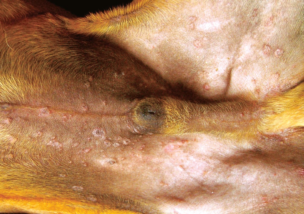
Picture 4 – Bacterial folliculitis: note epidermal papules and collarets on the abdomen and medial thighs
Papules in the abdominal area, in particular, are also occasionally observed during sarcoptic mange and food allergy. Crusts near mucocutaneous junctions, typical of mucocutaneous pyoderma, may mimic certain autoimmune diseases such as lupus erythematosus discoides.
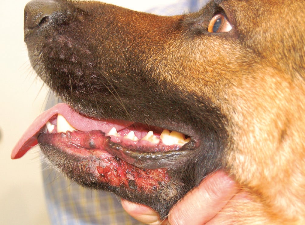
Picture 5 – Mucocutaneous perilabial pyoderma in a German shepherd
Demodicosis or dermatophytosis, which affect hair follicles and cause focal alopecia, may mimic bacterial folliculitis, especially in short-haired dogs. Finally, epitheliotropic lymphoma may occur in an erythematous-desquamative form very similar to diffuse superficial pyoderma.
Diagnosis
In cases of suspected pyoderma, the most useful diagnostic test is the cytological examination as it is easy to perform and is quick and inexpensive.
Sampling by apposition is obtained by gently pressing the slide directly on the lesion and is particularly suitable for sampling open and exposed areas such as exudative areas, open pustules or crusts raised with a small needle.
 For bacterial overgrowth and intertrigo, sampling using transparent adhesive tape (scotch test) is more indicated, which is then stained, as if it were a slide with Romanowsky-type staining (Diff-Quik, Emacolor).
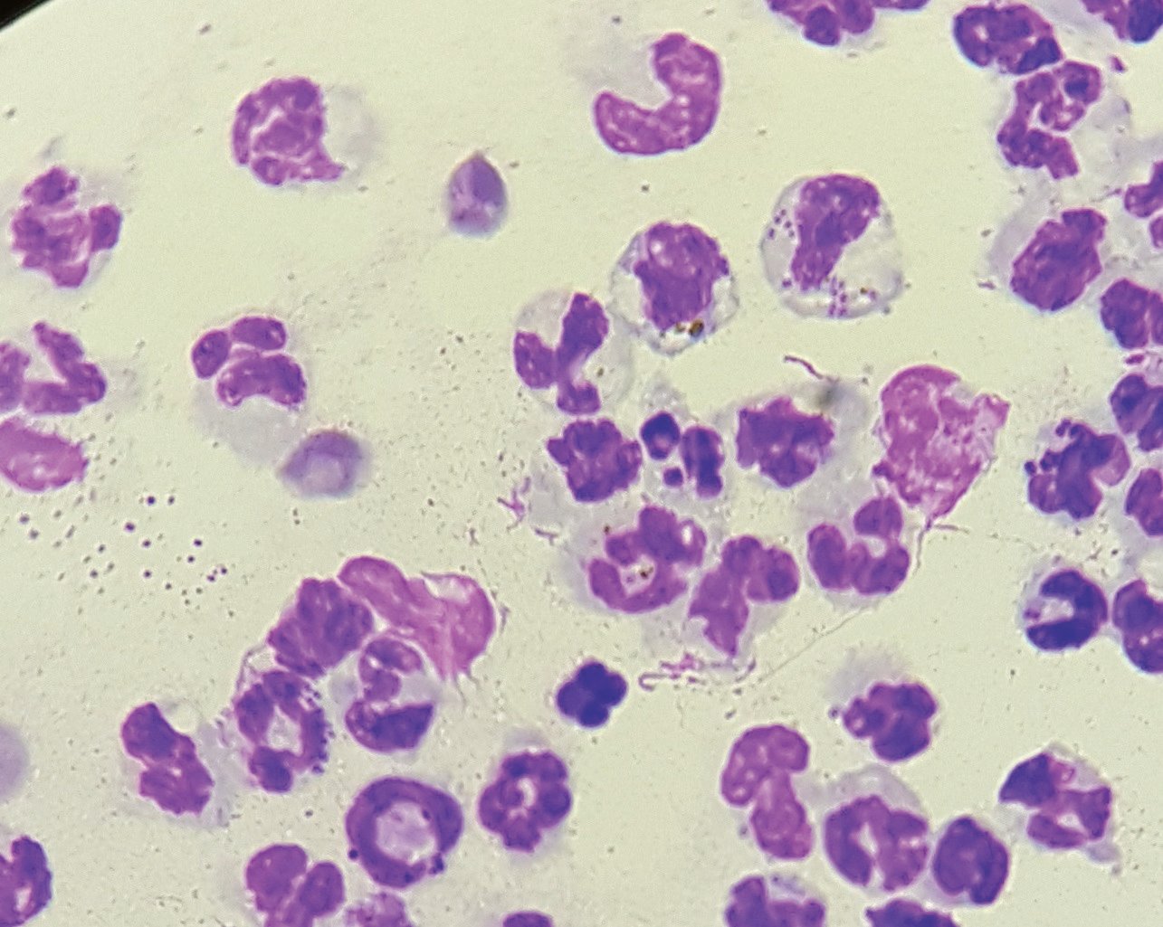
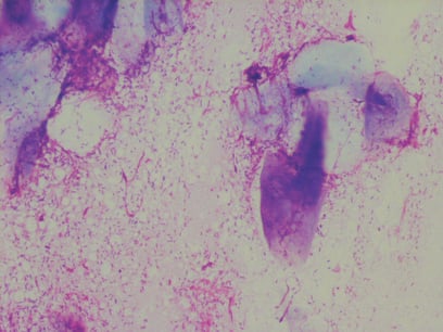
Picture 6 – Cytological examination showing the presence of intracellular bacteria in neutrophils, indicative of pyoderma
At the cytological examination, it is important to assess the presence of the bacteria and their localisation: in the case of true infections, these are observed within the inflammatory cells (Pic. 6), especially in the neutrophilic granulocytes and sometimes in the macrophages. If, on the other hand, they are only found at extracellular sites (Pic. 7), it is more likely to be bacterial overgrowth.

Picture 7 – Cytologic examination showing the presence of extracellular bacteria, indicative of bacterial overgrowth